
Görselleştirme : Verilerinizi Bilin bölümünde keşfedin
Açıklama :
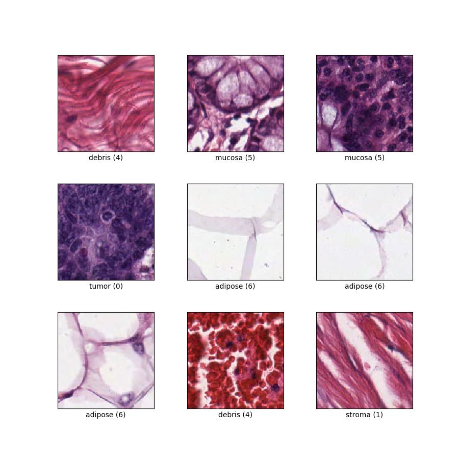
Kolorektal kanser histolojisinde dokuların sınıflandırılması. Her örnek, 8 sınıftan birinin 150 x 150 x 3 RGB görüntüsüdür.
Ana sayfa : https://zenodo.org/record/53169#.XGZemKwzbmG
Kaynak kodu :
tfds.image_classification.ColorectalHistologySürümler :
-
2.0.0(varsayılan): Yeni bölünmüş API ( https://tensorflow.org/datasets/splits )
-
İndirme boyutu :
246.14 MiBVeri kümesi boyutu :
179.23 MiBOtomatik önbelleğe alınmış ( belgeler ): Yalnızca
shuffle_files=False(train) olduğundaBölünmeler :
| Bölmek | Örnekler |
|---|---|
'train' | 5.000 |
- Özellik yapısı :
FeaturesDict({
    'filename': Text(shape=(), dtype=string),
    'image': Image(shape=(150, 150, 3), dtype=uint8),
    'label': ClassLabel(shape=(), dtype=int64, num_classes=8),
})
- Özellik belgeleri :
| Özellik | Sınıf | Şekil | Dtipi | Tanım |
|---|---|---|---|---|
| ÖzelliklerDict | ||||
| dosya adı | Metin | sicim | ||
| görüntü | Resim | (150, 150, 3) | uint8 | |
| etiket | SınıfEtiketi | int64 | Sekiz sınıf: 0: 'tümör epitelyumu', 1: 'basit stroma', 2: 'karmaşık stroma' (tek tümör hücreleri ve/veya tek bağışıklık hücrelerini içeren stroma), 3: 'bağışıklık hücresi kümeleri', 4: 'döküntü ve mukus', 5: 'mukozal bezler', 6: 'yağ dokusu' ve 7: 'arka plan'. |
Denetlenen anahtarlar (
as_supervisedbelgesine bakın):('image', 'label')Şekil ( tfds.show_examples ):
- Örnekler ( tfds.as_dataframe ):
- Alıntı :
@article{kather2016multi,
  title={Multi-class texture analysis in colorectal cancer histology},
  author={Kather, Jakob Nikolas and Weis, Cleo-Aron and Bianconi, Francesco and Melchers, Susanne M and Schad, Lothar R and Gaiser, Timo and Marx, Alexander and Z{"o}llner, Frank Gerrit},
  journal={Scientific reports},
  volume={6},
  pages={27988},
  year={2016},
  publisher={Nature Publishing Group}
}