
- Descripción :
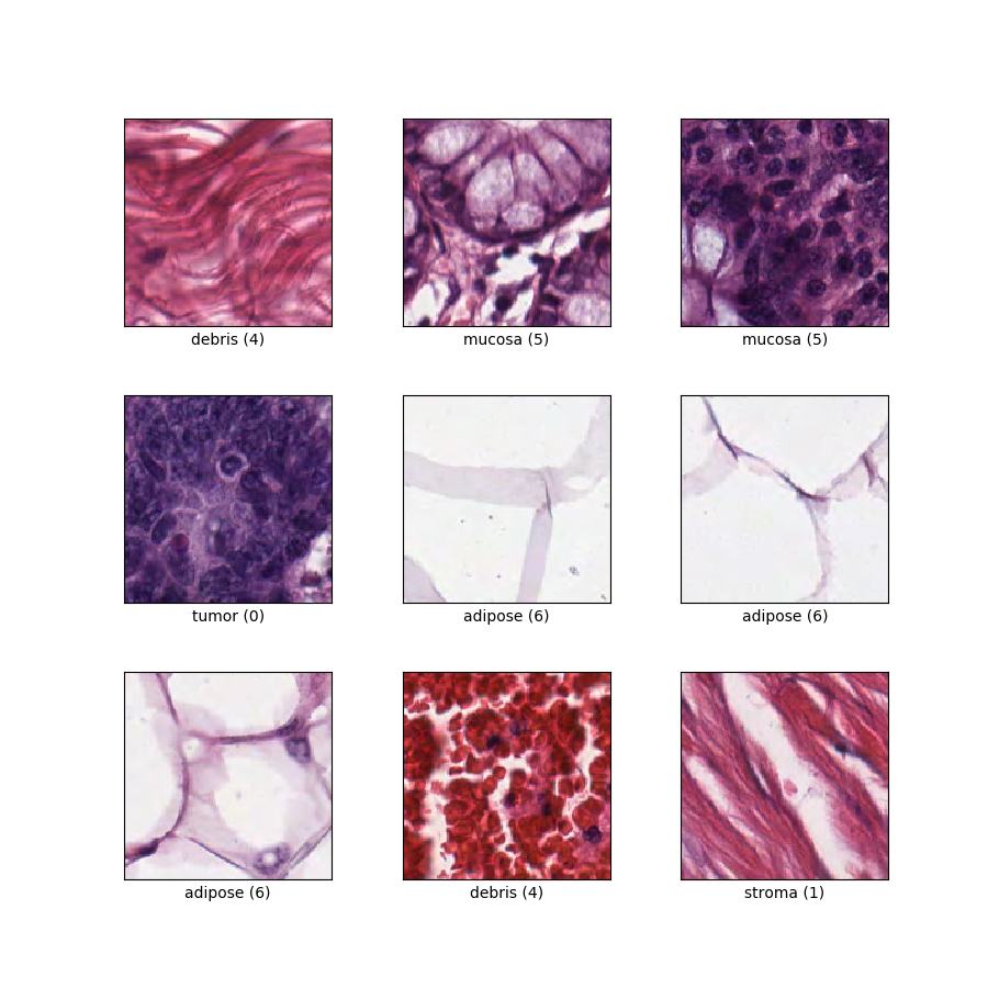
Clasificación de texturas en histología del cáncer colorrectal. Cada ejemplo es una imagen RGB de 150 x 150 x 3 de una de 8 clases.
Página de inicio : https://zenodo.org/record/53169#.XGZemKwzbmG
Código fuente :
tfds.image_classification.ColorectalHistologyVersiones :
-
2.0.0(predeterminado): Nueva API dividida ( https://tensorflow.org/datasets/splits )
-
Tamaño de descarga :
246.14 MiBTamaño del conjunto de datos :
179.23 MiBAlmacenamiento en caché automático ( documentación ): solo cuando
shuffle_files=False(entrenamiento)Divisiones :
| Dividir | Ejemplos |
|---|---|
'train' | 5.000 |
- Estructura de características :
FeaturesDict({
    'filename': Text(shape=(), dtype=string),
    'image': Image(shape=(150, 150, 3), dtype=uint8),
    'label': ClassLabel(shape=(), dtype=int64, num_classes=8),
})
- Documentación de funciones :
| Característica | Clase | Forma | tipo D | Descripción |
|---|---|---|---|---|
| FuncionesDict | ||||
| Nombre del archivo | Texto | cadena | ||
| imagen | Imagen | (150, 150, 3) | uint8 | |
| etiqueta | Etiqueta de clase | int64 | Ocho clases: 0: 'epitelio tumoral', 1: 'estroma simple', 2: 'estroma complejo' (estroma que contiene células tumorales individuales y/o células inmunes individuales), 3: 'conglomerados de células inmunes', 4: 'desechos y moco', 5: 'glándulas mucosas', 6: 'tejido adiposo' y 7: 'fondo'. |
Claves supervisadas (Ver documento
as_supervised):('image', 'label')Figura ( tfds.show_examples ):
- Ejemplos ( tfds.as_dataframe ):
- Citación :
@article{kather2016multi,
  title={Multi-class texture analysis in colorectal cancer histology},
  author={Kather, Jakob Nikolas and Weis, Cleo-Aron and Bianconi, Francesco and Melchers, Susanne M and Schad, Lothar R and Gaiser, Timo and Marx, Alexander and Z{"o}llner, Frank Gerrit},
  journal={Scientific reports},
  volume={6},
  pages={27988},
  year={2016},
  publisher={Nature Publishing Group}
}